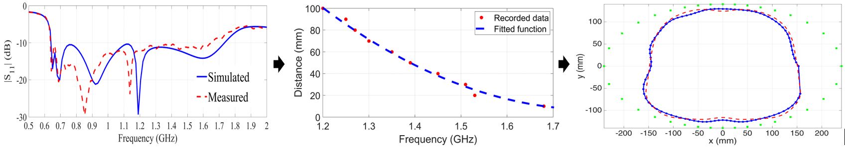
The performance of microwave imaging systems can be significantly improved by incorporating boundaries of the imaged object as a priori information. While it might be possible to manually measure that boundary in a controlled laboratory environment, this cannot be achieved in the clinical environment due to the impracticality of such a measurement in addition to the effect of the natural subject’s movement. In this paper, a method for boundary identification at the same time of imaging and using the same data captured for imaging is presented. The method is based on the relation between the imaging antennas’ resonant frequency and the location of the imaged object. The imaging antennas’ resonant frequency shifts when that antenna faces an imaging object, which is effectively a lossy dielectric in biomedical application, and that shift depends on the distance between the antenna and the object. The proposed technique is quite fast in scanning, computation, and image creation as it does not need additional devices for accurate boundary estimation. The method is tested via simulations and human trials using a torso imaging system. The collected data across the band 0.75-1.75 GHz using a 12-element antenna array, which is extended to 24 virtual elements, enclosing the human torso are processed to successfully estimate the torso boundary in a more accurate way than other methods. The included results in imaging a lung cancer case indicate that the accurate detection of the torso boundary improves microwave images.
Link to the paper